Examination of wound dressings
A wide variety of perspectives are possible on the product group of wound dressings. In the regulatory context, they can be medical devices or also medicinal products. Wound dressings can be classified differently in terms of their risk. In addition, they can be of a purely physical nature or have a pharmacological, immunological or metabolic effect.
At proderm, we address the versatility of wound dressings with a similar diversity of testing methods. In this respect, we have a comprehensive test portfolio for the investigation of wound dressings. We would like to briefly introduce the different approaches in the following.
Overview of the models
|
Suction blister model
In the suction blister model, wounds are created by means of negative pressure. Detailed information on the suction blister model is available in a case study that shows the successful use of the method in the examination of a wound healing cream.
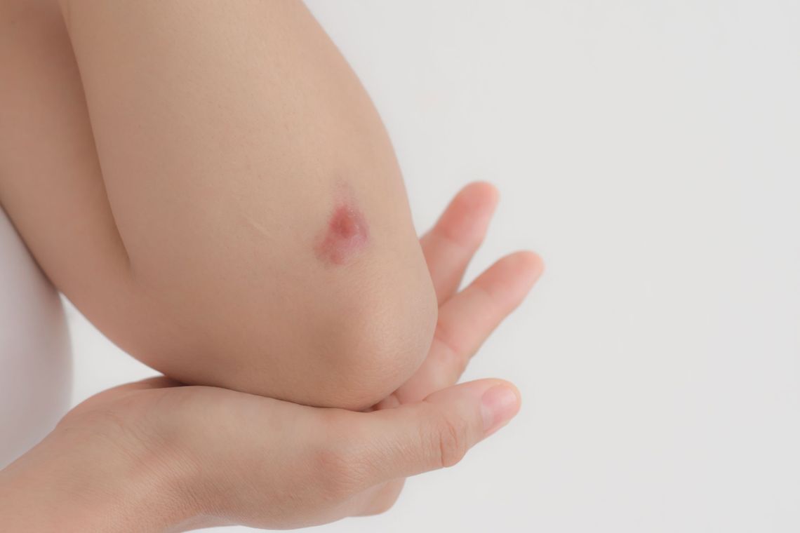
Abrasive wound model
Superficial wounds of about 2 cm² are obtained by standardised abrasion. The generated wounds have a close resemblance to real, everyday wounds. This procedure is not painful for the volunteer.
Laser model
The laser model is used to perform epidermal ablations with a laser. The depth of the wound between the upper epidermal layers to deeper wounds can be selected and created with great reproducibility. Local anaesthesia is required before creating the wound.
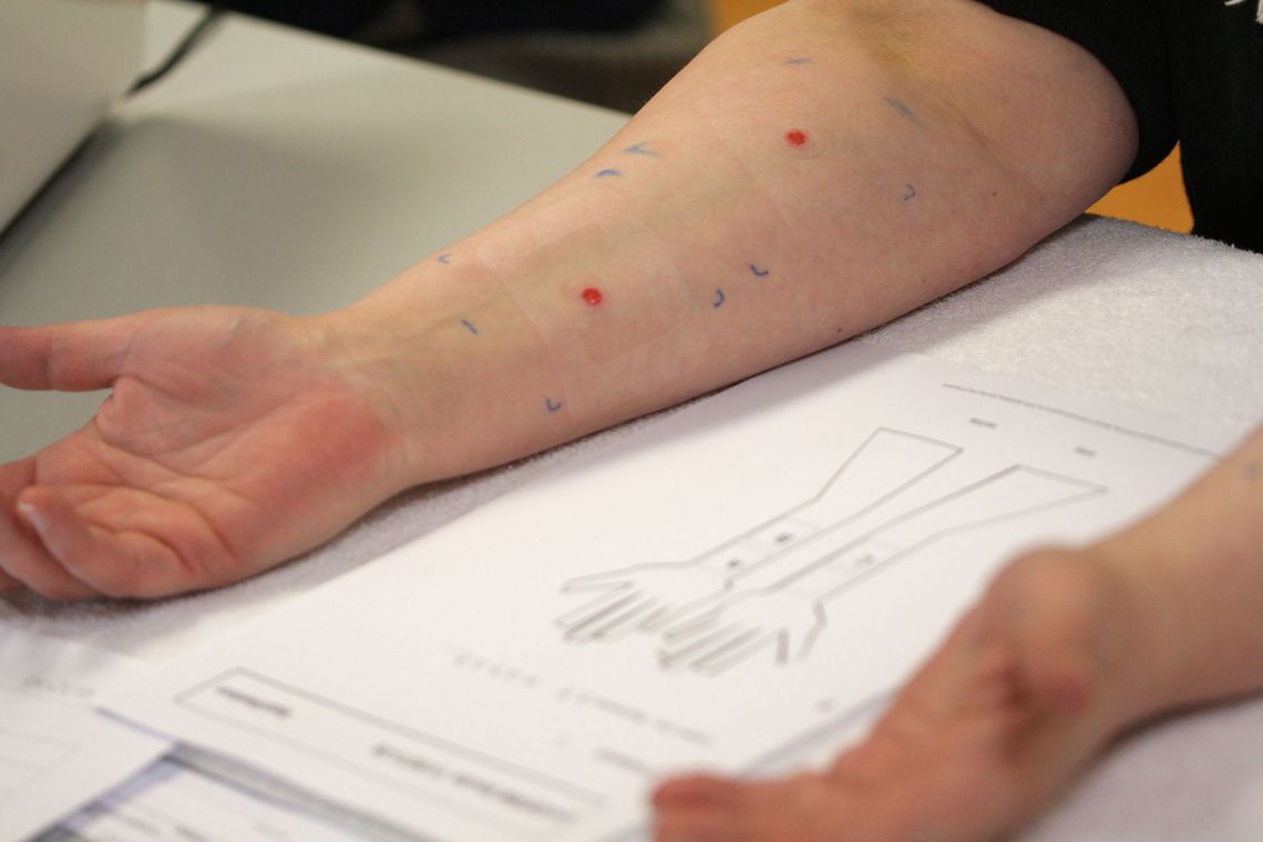
Scratch test
This model is preferably used when products for the treatment of superficial wounds are to be examined. With the help of a medical lancet, the skin is first damaged. During and after the 3 x 24-hour application phase, the wound is visually assessed.
Parameters at a glance
|
Adhesive force
Adhesive materials can be investigated with regard to their adhesive properties and peel off force required for removal. Adhesive material is applied on the back and removed with a universal test machine and a force meter.
Wound condition
We offer a wide variety of methods to examine the condition of the wound. For example, we use confocal microscopy to create images of different skin levels. We assess the skin barrier in the wound area by determining the transepidermal water loss. We can also determine the temperature and blood supply to the skin and take highly standardized clinical photos of the wound over time.
Wound healing
We can determine wound healing with instrumental and subjective procedures. These include, for example, visual assessment by trained investigators and study participants or examination by means of chromameter.
Evaluation of dressing
We can determine wound healing with instrumental and subjective procedures. These include, for example, visual assessment by trained investigators and study participants or examination by means of chromameter.
Contact
We will be happy to answer any questions you may have about the examinations. Please contact our medical team.